
Baker Cyst on MRI: Imaging Findings
A Baker's cyst on an MRI report is a fluid-filled bursa behind the knee, between the medial gastrocnemius and semimembranosus tendons, that connects to the joint. In adults it usually reflects a problem inside the knee, such as joint effusion, a meniscal tear or arthritis, rather than a separate disease.
Common Symptoms
- Palpable swelling or fullness in the back of the knee
- Dull aching discomfort when fully bending or extending the knee
- Tightness or pressure sensation behind the knee after activity
- Increased swelling after prolonged standing or walking
- Occasional popliteal cyst rupture causing sudden calf pain and swelling
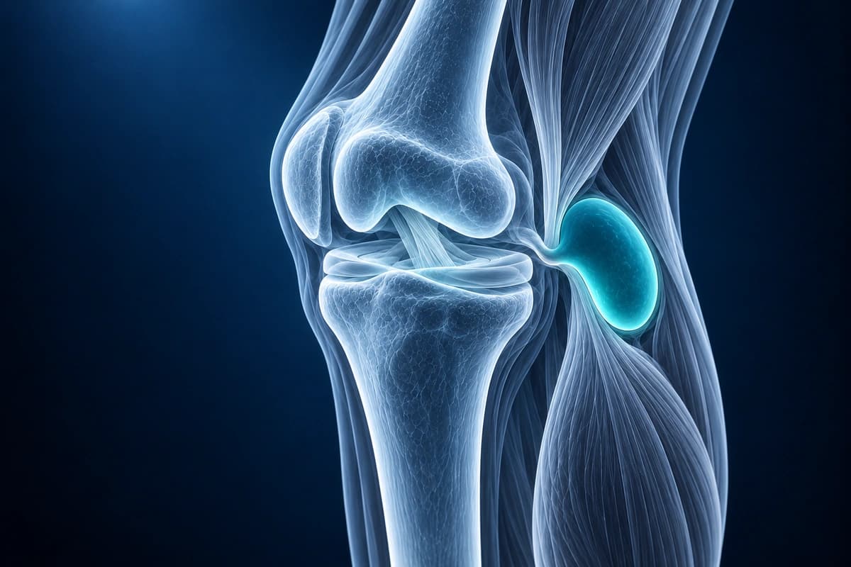
What your report says: “Baker's cyst” or “popliteal cyst”
Report wording you may see
- “Small popliteal (Baker's) cyst”
- “Distended gastrocnemius-semimembranosus bursa”
- “Ruptured Baker's cyst with fluid tracking into the calf”
- “Septated popliteal cyst with internal debris”
What it does and doesn't mean
- It is common. In one MRI study of older adults with knee osteoarthritis, a popliteal cyst was found in about a third of those with knee pain and in over a quarter of those without pain.
- It goes with effusion, meniscal tears and degenerative change: the more of these a knee has, the more likely a cyst is.
- “Ruptured” or “leaking” means fluid has escaped into the calf, which can cause sudden calf pain and swelling that looks like a blood clot.
When it tends to matter
Because the cyst is usually secondary, care tends to focus on the joint problem behind it. New calf swelling should be checked by a doctor promptly, as a ruptured cyst and a blood clot can look alike.
Questions for your doctor
- How large is the cyst, and does it connect to the joint?
- What inside my knee is likely feeding it: effusion, a meniscal tear or arthritis?
- Is there any sign of rupture or leakage into the calf?
- If my calf swells suddenly, what should I do?
A Baker's cyst (popliteal cyst) is a fluid-filled swelling behind the knee that typically communicates with the joint through a valvular mechanism between the semimembranosus tendon and the medial gastrocnemius. Baker's cysts are often secondary to intra-articular pathology such as meniscal tears, cartilage damage, or inflammatory arthritis.
What We Look For on Imaging
- Well-defined fluid collection in the posteromedial popliteal fossa
- Communication between the joint cavity and the cyst via the gastrocnemius-semimembranosus bursa
- High T2 signal indicating fluid content on MRI
- Internal debris or septations suggesting chronicity or hemorrhage
- Associated intra-articular pathology (meniscal tear, cartilage defect, effusion)
- Signs of rupture: fluid tracking along calf fascial planes
Frequently Asked Questions
Can AI detect a ruptured Baker's cyst on MRI?
A ruptured Baker's cyst can leave fluid tracking into the calf and resemble a blood clot, so new calf swelling needs prompt assessment. AI cannot exclude a clot.
What MRI sequences best visualize a Baker's cyst?
Sagittal and axial T2-weighted fat-saturated sequences provide the clearest delineation of cyst size, septations, and any internal debris.
Does a Baker's cyst always require treatment?
Most Baker's cysts are secondary to intra-articular pathology such as meniscus tears or osteoarthritis, and resolve when the underlying cause is treated. Isolated aspiration has high recurrence rates.
Sources
- Fritschy D, Fasel J, Imbert JC, et al. The popliteal cyst. Knee Surg Sports Traumatol Arthrosc. 2006;14(7):623-628.
- Miller TT, Staron RB, Koenigsberg T, et al. MR imaging of Baker cysts: association with internal derangement, effusion, and degenerative arthropathy. Radiology. 1996;201(1):247-250.
- Herman AM, Marzo JM. Popliteal cysts: a current review. Orthopedics. 2014;37(8):e678-e684.
- Hill CL, Gale DG, Chaisson CE, et al. Knee effusions, popliteal cysts, and synovial thickening: association with knee pain in osteoarthritis. J Rheumatol. 2001;28(6):1330-1337.
Related Articles
Your knee MRI report mentions joint effusion? Learn what a small, moderate or large effusion means, what can cause fluid in the knee and what to ask your doctor.
Learn to understand your knee MRI report, common sequences, and what key findings mean for your diagnosis.
Understand your imaging before your next appointment
MRI, X-ray or CT from a CD, folder or ZIP. A PDF report or photos of the films work too.
Free to open and preview, no account needed. An optional AI report from $10 explains your images in plain language, with questions for your doctor.
Your raw files stay on this device. Only rendered images and minimal context are sent when you request an AI report.
Medical Disclaimer: This page is for informational and educational purposes only. It does not constitute medical advice, diagnosis, or treatment. AI-generated analysis may contain errors. Always consult a qualified healthcare professional for medical decisions. Full Disclaimer