
3D DICOM Viewer, MPR, and Volume Rendering
Understand 3D DICOM viewing, MPR reconstruction, and browser volume rendering for MRI, CT, and CBCT scans, including privacy and scan compatibility limits.
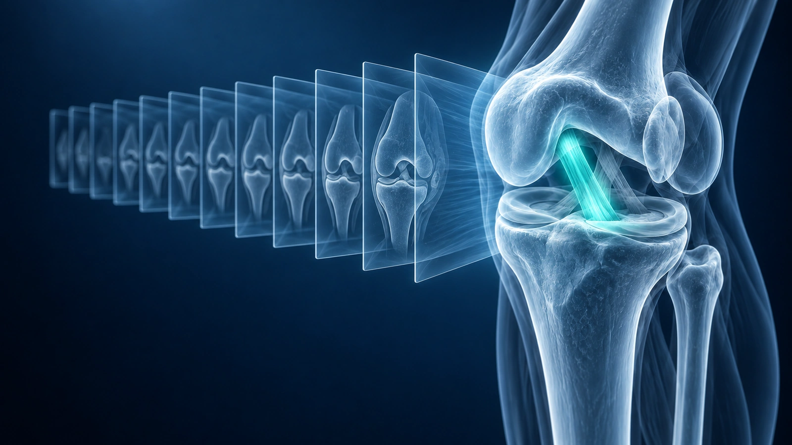
A 3D DICOM viewer turns a compatible stack of medical image slices into a volume that can be rotated, reconstructed, or inspected in multiple planes. The same idea applies to MRI, CT, and dental CBCT, but scan quality and acquisition protocol decide what the 3D view can actually show.
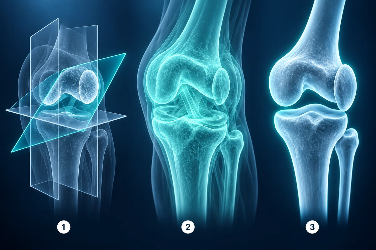
The main terms are MPR, volume rendering, and surface-style 3D rendering. They sound similar, but they answer different viewing questions. Understanding the difference helps you avoid over-reading a pretty 3D view as if it were a diagnosis.
MPR vs 3D Rendering vs Volume Rendering
| Mode | What it shows | Best for | Common limit |
|---|---|---|---|
| MPR | Rebuilt axial, sagittal, coronal, or oblique slices | Careful cross-plane review | Thick or uneven slices reduce detail |
| Volume rendering | Transparent projection through the whole voxel volume | Anatomy orientation and explaining spatial relationships | Preset choices can hide subtle findings |
| Surface-style 3D | A clearer outer structure or tissue boundary view | Bone, joint, implant, and broad anatomy visualization | Small internal soft-tissue findings may disappear |
Which Scans Work Best for 3D
3D viewing works best when the study has many closely spaced slices, consistent orientation, accurate spacing metadata, and enough coverage across the anatomy. CT and CBCT often produce strong 3D views because the data is commonly acquired as a volume. MRI can work well too, especially with dedicated 3D or thin-slice sequences.
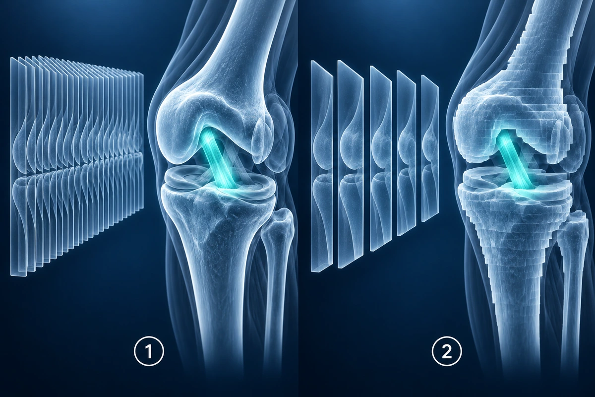
A single X-ray, PDF report, screenshot, small localizer, or sparse series cannot become a meaningful 3D volume. Those files may still be useful, but they should be viewed in their original 2D form.
Browser-Based 3D DICOM Viewing
Modern browsers can decode supported DICOM files, build image stacks, and render 3D views using WebGL or related graphics APIs. This makes quick viewing possible without a desktop PACS workstation or old hospital CD viewer.
For viewing, raw files can stay in the browser. When optional AI explanation is requested, the product uses rendered images and minimal context rather than turning the server into a medical file store.
Open the MRI viewer with 3D supportWhy MPR Matters Clinically
MPR is often more clinically useful than a dramatic 3D render because it preserves a slice-like view. It lets you inspect anatomy in a plane that was not originally acquired, such as checking a joint line, fracture orientation, dental implant corridor, or relationship between a lesion and nearby structures.
Even then, MPR depends on source data. If the original slices are thick, far apart, or affected by motion, reconstructed planes can look blocky or misleading. The radiology report and original diagnostic sequences remain the reference point.
3D Knee MRI as a Practical Example
In a compatible knee MRI, 3D rendering can help you understand the relationship between femur, tibia, patella, joint space, fluid, and soft-tissue structures. It is especially useful before reading sagittal and coronal slices because it gives spatial context.
It is less reliable for subtle meniscus signal, partial ligament tears, cartilage fissures, marrow edema, and postoperative changes. Those findings need slice review and clinician interpretation.
Read the knee-specific 3D MRI render guideCompatibility Checklist
- Enough slices to cover the anatomy as a volume
- Consistent slice spacing and orientation metadata
- Supported DICOM transfer syntax and pixel depth
- Device memory sufficient for the selected study
- A rendering preset that matches the anatomy you want to inspect
Key Takeaways
- MPR rebuilds slice planes; volume rendering creates a rotatable projection
- CT and CBCT often render well because they are commonly volumetric
- MRI 3D quality depends heavily on sequence type, slice spacing, and motion
- 3D views help orientation but can hide subtle diagnostic findings
- Use 3D alongside original images, reports, and clinician review
Frequently Asked Questions
What is a 3D DICOM viewer?
It is a viewer that can reconstruct compatible DICOM slices as a volume, allowing MPR, rotation, or volume rendering depending on scan data and device capability.
Is MPR the same as 3D rendering?
No. MPR reconstructs flat planes through a volume. 3D rendering projects the volume into a rotatable view. Both depend on the same underlying scan data.
Can every MRI be shown in 3D?
No. Some MRI studies are too sparse, thick-sliced, motion-affected, or limited in coverage to produce a useful 3D view. They can still be valuable in the 2D viewer.
Related Articles
Learn how compatible knee MRI DICOM slices become a private 3D joint view, what MPR means, and what 3D rendering can and cannot show.
Compare browser-based DICOM viewing, cloud upload viewers, and desktop apps so you can open MRI, CT, and X-ray files with the right privacy tradeoffs.
Step-by-step guide to opening DICOM files (.dcm) from your hospital MRI/CT/X-ray CD or USB on Mac, Windows, iPhone, and Android — no software install required.
Related Conditions
Ready to analyze your imaging? Upload your MRI or X-ray for AI-powered analysis
Upload your MRI or X-ray DICOM files for private, AI-powered analysis. 4 models analyze independently — raw scan files stay in your browser.
Start AnalysisMedical Disclaimer: This page is for informational and educational purposes only. It does not constitute medical advice, diagnosis, or treatment. AI-generated analysis may contain errors. Always consult a qualified healthcare professional for medical decisions. Full Disclaimer