
Knee MCL and LCL Tear MRI Signs: Ligament Sprain Grades
The medial collateral ligament (MCL) and lateral collateral ligament (LCL) are the primary stabilizers against valgus and varus forces at the knee. MCL injuries are far more common, typically resulting from contact sports or direct blows to the lateral knee, while LCL tears occur from varus stress or hyperextension mechanisms.
Common Symptoms
- Pain and tenderness directly over the medial or lateral knee joint line
- Valgus or varus instability during side-to-side stress testing
- Swelling localized to the inner or outer aspect of the knee
- Difficulty walking, especially on uneven surfaces
- Bruising along the medial or lateral knee following impact
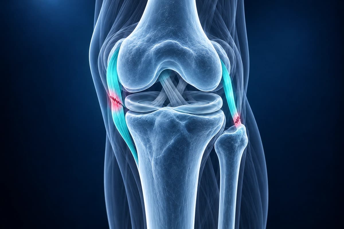
A useful knee MRI review does not stop at the ligament. It should also check for compartment-specific bone bruise patterns, nearby meniscus injury, and combined cruciate ligament trauma such as an ACL tear. Those associated findings often explain why a grade 1 or grade 2 sprain still feels unstable.
What We Look For on Imaging
- High signal intensity within or surrounding the MCL or LCL on coronal MRI
- Ligament thickening, waviness, or complete fiber discontinuity
- Periligamentous edema and subcutaneous soft tissue swelling
- Grade classification based on fiber integrity and gapping
- Associated bone bruise pattern on the opposite compartment
- Concomitant meniscal tears or cruciate ligament injuries
Frequently Asked Questions
How does AI grade MCL and LCL tears on MRI?
Grade I sprains retain intact fibers, grade II tears disrupt some fibers, and grade III tears are complete; examination assesses stability. AI cannot assign a clinical injury grade.
What MRI sequences are best for collateral ligament evaluation?
Coronal PD fat-saturated sequences provide the clearest view of the MCL and LCL along their full length.
When does a collateral ligament tear require surgery?
Isolated Grade I–II MCL tears typically heal with conservative management. Complete Grade III tears, especially of the LCL or those associated with posterolateral corner injuries, often require surgical repair or reconstruction.
Sources
- Hughston JC, Andrews JR, Cross MJ, Moschi A. Classification of knee ligament instabilities. Part I. The medial compartment and cruciate ligaments. J Bone Joint Surg Am. 1976;58(2):159-172.
- Hughston JC, Andrews JR, Cross MJ, Moschi A. Classification of knee ligament instabilities. Part II. The lateral compartment. J Bone Joint Surg Am. 1976;58(2):173-179.
- Fetto JF, Marshall JL. Medial collateral ligament injuries of the knee: a rationale for treatment. Clin Orthop Relat Res. 1978;(132):206-218.
- Schweitzer ME, Tran D, Deely DM, Hume EL. Medial collateral ligament injuries: evaluation of multiple signs, prevalence and location of associated bone bruises, and assessment with MR imaging. Radiology. 1995;194(3):825-829.
- American Academy of Orthopaedic Surgeons. Collateral Ligament Injuries. OrthoInfo.
Related Articles
Understand ACL tear grading from mild sprains to complete ruptures, MRI findings for each grade, and treatment implications.
Understand the differences between ACL and MCL tears, injury mechanisms, symptoms, MRI findings, and treatment approaches.
Learn to understand your knee MRI report, common sequences, and what key findings mean for your diagnosis.
Understand your imaging before your next appointment
MRI, X-ray or CT from a CD, folder or ZIP. A PDF report or photos of the films work too.
Free to open and preview, no account needed. An optional AI report from $10 explains your images in plain language, with questions for your doctor.
Your raw files stay on this device. Only rendered images and minimal context are sent when you request an AI report.
Medical Disclaimer: This page is for informational and educational purposes only. It does not constitute medical advice, diagnosis, or treatment. AI-generated analysis may contain errors. Always consult a qualified healthcare professional for medical decisions. Full Disclaimer