
How to Read a Knee MRI: A Patient's Guide
Learn to understand your knee MRI report, common sequences, and what key findings mean for your diagnosis.
Key Takeaways
- Knee MRI uses three planes (sagittal, coronal, axial) and two main sequences (T1 and T2)
- Normal ligaments and menisci appear dark; bright signal within them suggests injury
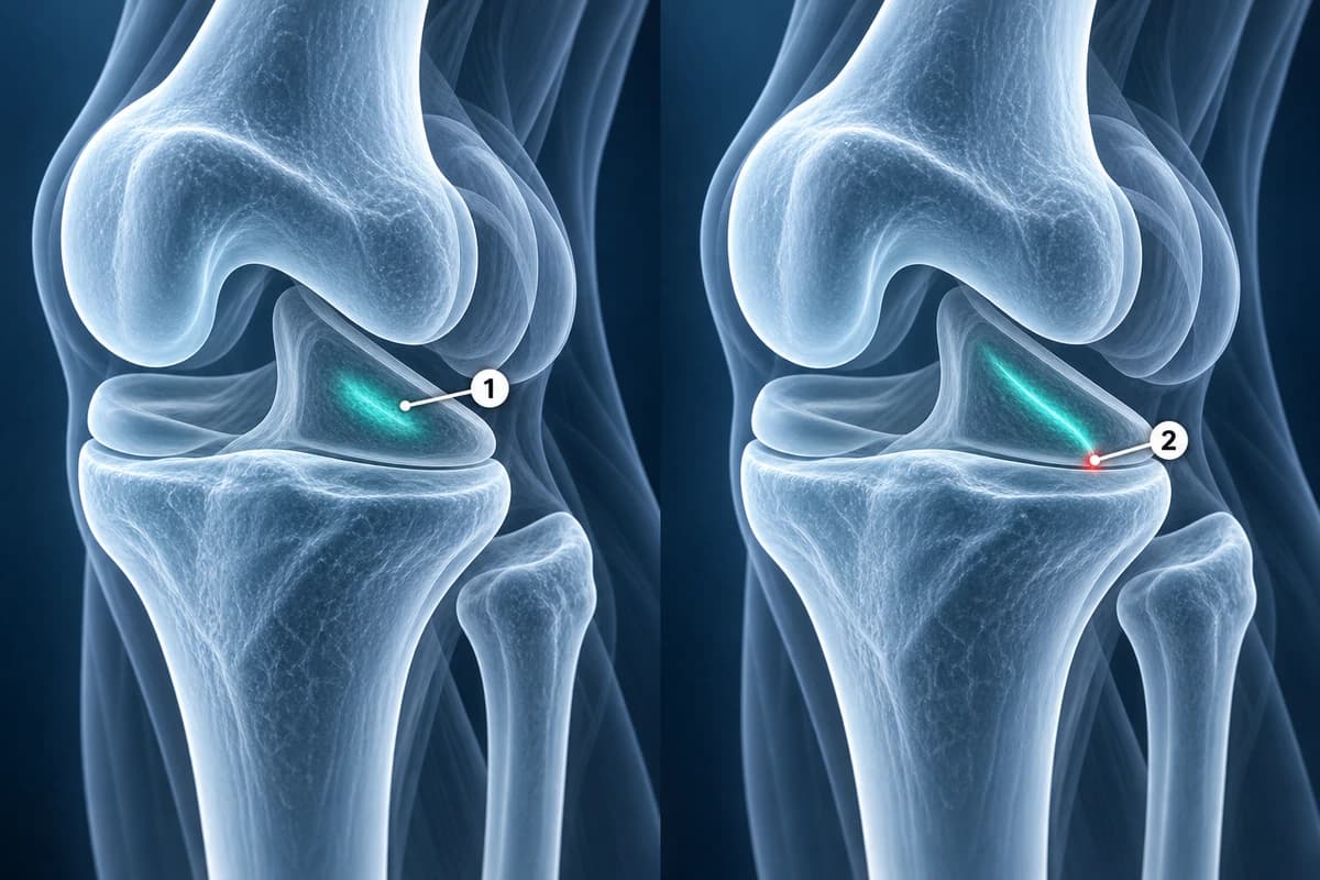
- Meniscal tears require abnormal signal to reach the articular surface — internal signal alone is not a tear
- Many MRI findings (mild degeneration, small effusions) are normal with aging and may not require treatment
- Bone marrow edema (bright on T2, dark on T1) indicates stress or contusion
- Always correlate MRI findings with symptoms — not every abnormality on MRI is clinically significant

Receiving a knee MRI can feel overwhelming — the images look like abstract art and the radiology report is full of unfamiliar terminology. But understanding the basics of knee MRI reading can help you have more informed conversations with your doctor and better understand your diagnosis. This guide breaks down what each structure looks like on MRI, how to distinguish normal from abnormal, and what common findings actually mean for your knee health.
MRI (magnetic resonance imaging) uses powerful magnets and radio waves to create detailed cross-sectional images of the knee without radiation. Unlike X-rays, which primarily show bone, MRI excels at visualizing soft tissues including ligaments, menisci, cartilage, and tendons. This makes it the gold standard imaging study for evaluating most knee problems.
MRI Basics: Views and Sequences
A knee MRI is typically acquired in three planes: sagittal (side view, dividing left from right), coronal (front view, dividing front from back), and axial (top-down slices). Each plane is best for evaluating specific structures. The sagittal view is ideal for the ACL, PCL, and menisci. The coronal view best shows the collateral ligaments and meniscal body. The axial view is used for the patella, patellar tendon, and cartilage under the kneecap.
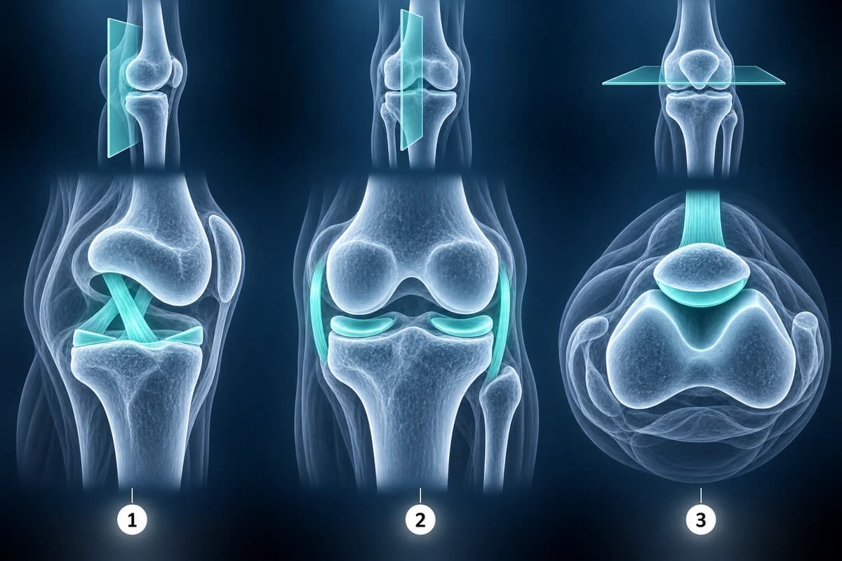
Two main sequence types appear in most knee MRIs: T1-weighted images show anatomy clearly with fat appearing bright and fluid appearing dark. T2-weighted (or proton density fat-saturated) images highlight pathology because fluid and inflammation appear bright white. Understanding this basic principle — that bright signal on T2 within a normally dark structure suggests injury — is the key to reading knee MRI.
The Anterior Cruciate Ligament (ACL)
The ACL is best seen on sagittal images as a dark, taut band running from the posterior femur to the anterior tibia. A normal ACL has uniform low signal (dark) with clearly defined parallel fibers. Signs of an ACL tear include complete absence of the ligament on expected slices, a wavy or lax appearance instead of a taut band, bright signal within the ligament substance indicating edema or hemorrhage, and abnormal orientation.
Secondary signs of ACL injury include bone bruising on the lateral femoral condyle and posterolateral tibial plateau, anterior tibial translation, and a deep lateral femoral notch sign. For a detailed breakdown of injury severity, see our guide on ACL tear grades.
Meniscus Evaluation
The menisci appear as dark triangular or bowtie-shaped structures on MRI. The medial meniscus is C-shaped and the lateral meniscus is more circular. On sagittal images, you see the anterior horn, body (which has a characteristic bowtie shape on two consecutive slices), and posterior horn. A normal meniscus is uniformly dark (low signal) on all sequences.
Meniscal tears are diagnosed when abnormal bright signal extends to the articular surface — that is, the signal touches at least one edge of the meniscus. Internal signal that does not reach the surface (grades 1 and 2) represents degeneration, not a true tear. A grade 3 signal that touches the surface is a definitive tear. For a complete overview of tear types and classifications, see our article on meniscus tear types.
Cartilage, Bone, and Fluid
In one study, knee bone bruises remained visible on MRI in 69% of patients at four months and 12% at twelve months. See MRI vs X-ray.
If you are using AI to understand report wording, keep the distinction clear: an AI explanation can help you prepare questions, while a radiology second opinion is a licensed medical interpretation. See the AI MRI explanation vs second opinion guide.
Frequently Asked Questions
Can I see a meniscus tear on MRI myself?
Yes, with basic knowledge you can often identify meniscal tears. Look for bright signal within the normally dark meniscus that extends to the top or bottom surface on sagittal or coronal images. The tear appears as a bright line or area within the dark triangle. However, subtle tears and complex tear patterns can be difficult to detect without training, and only a radiologist's report is a diagnosis. An AI explanation can help you understand the report wording and prepare questions for your doctor.
What does increased signal mean on my MRI report?
Increased signal means an area appears brighter than expected on a particular MRI sequence. In structures that should be dark (like ligaments and menisci), increased signal suggests damage, inflammation, or degeneration. In bone (on T2/STIR sequences), increased signal indicates edema or bruising. The clinical significance depends on the location, extent, and sequence on which the signal change is seen.
How accurate is knee MRI for detecting tears?
A meta-analysis estimated MRI sensitivity and specificity at 87% and 93% for ACL tears, 89% and 88% for medial meniscal tears, and 78% and 95% for lateral meniscal tears.
Do I need contrast dye for a knee MRI?
Most routine knee MRIs do not require intravenous contrast. Standard non-contrast sequences are excellent for evaluating ligaments, menisci, cartilage, and bone. Contrast (gadolinium) may be used when evaluating for tumors, infections, or synovial conditions. MR arthrography is sometimes used for evaluating subtle cartilage defects but is rarely needed for routine knee evaluation.
Sources
- Stoller DW, Martin C, Crues JV 3rd, et al. Meniscal tears: pathologic correlation with MR imaging. Radiology. 1987;163(3):731-735.
- Phelan N, Rowland P, Galvin R, O'Byrne JM. A systematic review and meta-analysis of the diagnostic accuracy of MRI for suspected ACL and meniscal tears of the knee. Knee Surg Sports Traumatol Arthrosc. 2016;24(5):1525-1539.
- Sanders TG, Medynski MA, Feller JF, Lawhorn KW. Bone contusion patterns of the knee at MR imaging: footprint of the mechanism of injury. Radiographics. 2000;20 Spec No:S135-S151.
- Bretlau T, Tuxøe J, Larsen L, et al. Bone bruise in the acutely injured knee. Knee Surg Sports Traumatol Arthrosc. 2002;10(2):96-101.
Related Articles
Understand ACL tear grading from mild sprains to complete ruptures, MRI findings for each grade, and treatment implications.
Your MRI names a meniscus tear pattern? Learn what horizontal, radial, bucket-handle and complex tears mean, why the zone matters and how each is treated.
Compare MRI and X-ray imaging, understand when each is appropriate, radiation considerations, cost, and diagnostic capabilities.
Related Conditions
Understand your imaging before your next appointment
MRI, X-ray or CT from a CD, folder or ZIP. A PDF report or photos of the films work too.
Free to open and preview, no account needed. An optional AI report from $10 explains your images in plain language, with questions for your doctor.
Your raw files stay on this device. Only rendered images and minimal context are sent when you request an AI report.
Medical Disclaimer: This page is for informational and educational purposes only. It does not constitute medical advice, diagnosis, or treatment. AI-generated analysis may contain errors. Always consult a qualified healthcare professional for medical decisions. Full Disclaimer