
Knee Patellar Dislocation MRI Signs: MPFL Tear and Instability
Patellar dislocation occurs when the patella displaces laterally out of the trochlear groove, most commonly during pivoting or twisting movements. It is frequently associated with medial patellofemoral ligament (MPFL) tears, trochlear dysplasia, and characteristic bone bruise patterns. AI can help explain these findings, but a clinician must assess the risk of another dislocation and discuss non-surgical or surgical treatment.
Common Symptoms
- Sudden lateral displacement of the kneecap during twisting or contact
- Acute medial peripatellar pain from medial patellofemoral ligament (MPFL) disruption
- Significant knee swelling developing rapidly after the event
- Apprehension and fear of re-dislocation with lateral patellar pressure
- Tenderness over the medial patellar border and femoral attachment of the MPFL
- Visible or palpable patellar malalignment in the acute setting
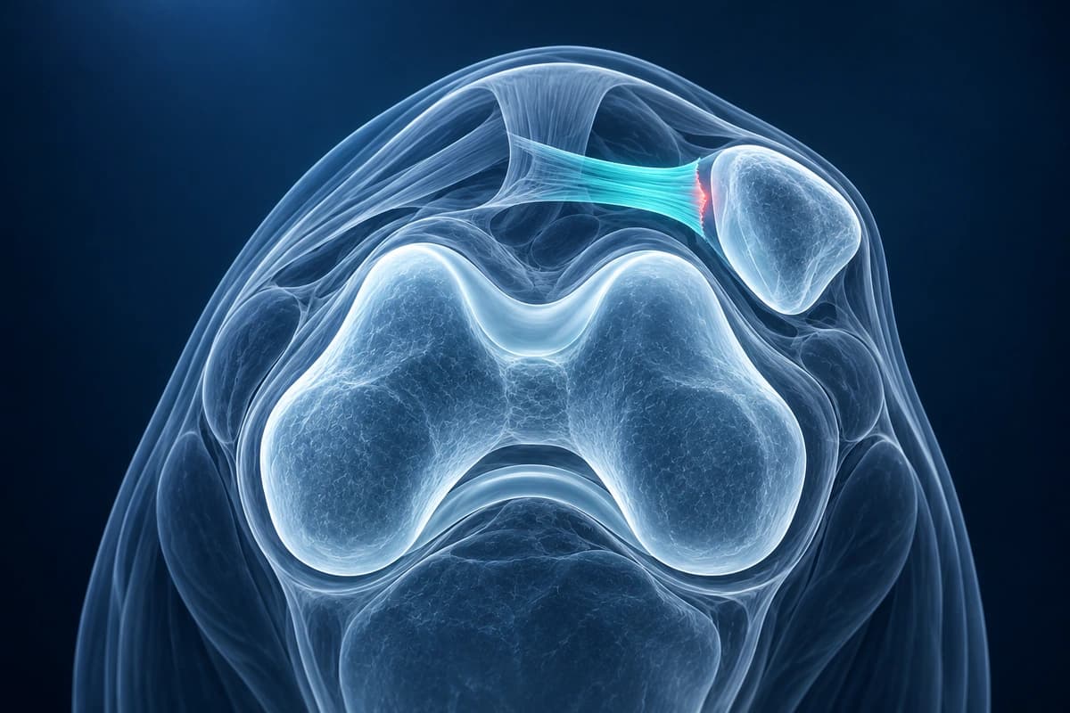
The highest-yield MRI questions are whether the MPFL is torn, whether there is a medial patellar facet or lateral femoral condyle bone bruise, and whether a loose osteochondral fragment is present. For broader knee scan orientation, compare this page with the knee MRI reading guide.
What We Look For on Imaging
- Bone bruise on the medial patellar facet and lateral femoral condyle
- MPFL tear or avulsion at the patellar or femoral attachment
- Trochlear dysplasia with shallow or flat trochlear groove
- Lateral patellar tilt or subluxation on axial images
- Osteochondral fracture fragments or loose bodies within the joint
- Joint effusion and hemarthrosis following acute dislocation
Frequently Asked Questions
Can AI detect MPFL tears after patellar dislocation?
MRI can show injury to the medial patellofemoral ligament after a kneecap dislocation, along with cartilage or bone injury. AI cannot confirm ligament integrity or decide treatment.
What MRI findings suggest high risk of recurrent patellar dislocation?
Structural risk factors including trochlear dysplasia, patella alta, and increased tibial tuberosity–trochlear groove (TT-TG) distance are associated with recurrent instability.
Are osteochondral injuries detectable after patellar dislocation?
Lateral femoral condyle and medial patellar facet osteochondral injuries occur in approximately 40% of first-time dislocations. An AI explanation can help you prepare questions for your clinician. It cannot confirm an injury or its grade on its own and is not a diagnosis.
Sources
- Diederichs G, Issever AS, Scheffler S. MR imaging of patellar instability: injury patterns and assessment of risk factors. Radiographics. 2010;30(4):961-981.
- Elias DA, White LM, Fithian DC. Acute lateral patellar dislocation at MR imaging: injury patterns of medial patellar soft-tissue restraints and osteochondral injuries of the inferomedial patella. Radiology. 2002;225(3):736-743.
- Dejour H, Walch G, Nove-Josserand L, Guier C. Factors of patellar instability: an anatomic radiographic study. Knee Surg Sports Traumatol Arthrosc. 1994;2(1):19-26.
- American Academy of Orthopaedic Surgeons. Unstable Kneecap (Patellar Instability). OrthoInfo.
Understand your imaging before your next appointment
MRI, X-ray or CT from a CD, folder or ZIP. A PDF report or photos of the films work too.
Free to open and preview, no account needed. An optional AI report from $10 explains your images in plain language, with questions for your doctor.
Your raw files stay on this device. Only rendered images and minimal context are sent when you request an AI report.
Medical Disclaimer: This page is for informational and educational purposes only. It does not constitute medical advice, diagnosis, or treatment. AI-generated analysis may contain errors. Always consult a qualified healthcare professional for medical decisions. Full Disclaimer