
PCL Tear on MRI: Imaging Findings
The posterior cruciate ligament (PCL) is the strongest ligament in the knee and is less commonly injured than the ACL. PCL injuries typically result from dashboard injuries, falls on a flexed knee, or hyperextension.
Common Symptoms
- Posterior knee pain following a dashboard-type impact or fall
- Posterior sag of the tibia visible compared to the uninjured leg
- Instability or giving way, particularly when descending stairs
- Aching discomfort at the back of the knee with prolonged sitting
- Swelling that may be milder than ACL injuries
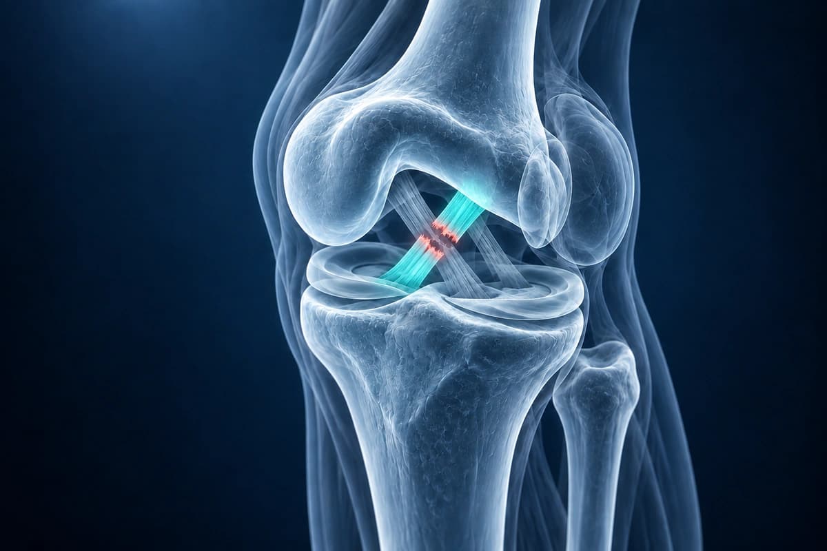
What We Look For on Imaging
- Increased signal intensity within PCL substance on sagittal MRI
- PCL fiber discontinuity or complete disruption
- Thickening and abnormal morphology of the PCL
- Bone bruise on anterior tibial plateau (dashboard injury pattern)
- Posterior tibial translation on sagittal images
- Associated posterolateral corner injury findings
Frequently Asked Questions
Can AI differentiate a partial from complete PCL tear on MRI?
Partial PCL tears retain some fibers, while complete tears disrupt the ligament; MRI and stability testing are assessed together. AI cannot determine whether the remaining ligament functions normally.
What MRI view is most important for PCL evaluation?
The sagittal plane is the primary view for PCL assessment, where the full course of the ligament from the femoral condyle to the tibial insertion is visible.
How accurate is AI at detecting PCL tears?
MRI helps assess PCL tears and associated injuries, alongside clinical stability tests. This service has no established clinical accuracy for PCL tear detection.
Sources
- Grover JS, Bassett LW, Gross ML, et al. Posterior cruciate ligament: MR imaging. Radiology. 1990;174(2):527-530.
- Sonin AH, Fitzgerald SW, Friedman H, et al. Posterior cruciate ligament injury: MR imaging diagnosis and patterns of injury. Radiology. 1994;190(2):455-458.
- Fanelli GC, Edson CJ. Posterior cruciate ligament injuries in trauma patients: Part II. Arthroscopy. 1995;11(5):526-529.
- American Academy of Orthopaedic Surgeons. Posterior Cruciate Ligament Injuries. OrthoInfo.
Understand your imaging before your next appointment
MRI, X-ray or CT from a CD, folder or ZIP. A PDF report or photos of the films work too.
Free to open and preview, no account needed. An optional AI report from $10 explains your images in plain language, with questions for your doctor.
Your raw files stay on this device. Only rendered images and minimal context are sent when you request an AI report.
Medical Disclaimer: This page is for informational and educational purposes only. It does not constitute medical advice, diagnosis, or treatment. AI-generated analysis may contain errors. Always consult a qualified healthcare professional for medical decisions. Full Disclaimer