
Common Symptoms
- Anterior knee pain that worsens with prolonged sitting (movie-goer's sign)
- Aching pain when ascending or descending stairs
- Crepitus or grinding felt under the kneecap during movement
- Diffuse peripatellar pain after running or squatting
- Occasional patellar swelling after intense activity
Chondromalacia: AI-Powered MRI Analysis
AI-powered chondromalacia imaging analysis on knee MRI. See how patellar cartilage softening and damage appear from early changes to full-thickness defects. 4 AI models analyze cartilage signal and morphology in parallel.
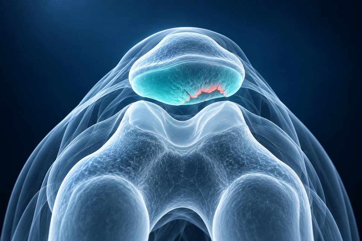
What your report says: chondromalacia patellae grade 1 to 4
Chondromalacia patellae means the cartilage on the back of the kneecap is softened or damaged. Reports grade it from 1 to 4: grade 1 is softening or swelling, grades 2 and 3 are fissures or defects of increasing size or depth, and grade 4 means the cartilage has worn through to the bone underneath.
Upload Knee ImagingReport wording you may see
- “Grade 2 chondromalacia patellae at the median ridge”
- “Chondral fissuring of the lateral patellar facet”
- “Patellar chondrosis” or “high-grade patellofemoral chondral loss”
- “Full-thickness cartilage defect with subchondral edema”
How the grading works
- The original Outerbridge scale was described at surgery. It separates grades 2 and 3 by the size of the damaged area, and grade 4 is erosion down to bone.
- The ICRS scale grades by depth instead: grade 2 involves less than half of the cartilage thickness and grade 3 more than half.
- Cartilage-sensitive MRI sequences detect most kneecap cartilage lesions; in a cadaver study an optimized sequence found 96% of them.
- Location words point to the spot: the medial and lateral facets and the median ridge are areas of the kneecap's underside, and the trochlea is the groove it glides in.
When it tends to matter
A grade alone does not decide treatment; your doctor weighs it with your symptoms, kneecap tracking and examination. Deep or full-thickness defects are the findings most often discussed in detail with an orthopaedic specialist.
Questions for your doctor
- Which grading scale does my report use, and is my grade based on depth or area?
- Which part of the kneecap or the trochlea is affected?
- Is the bone under the cartilage involved?
- Which exercises or activity changes make sense for this grade?
Chondromalacia patella refers to softening, fissuring, and erosion of the cartilage on the undersurface of the patella (kneecap). It is a common cause of anterior knee pain, particularly in young adults and athletes. MRI with cartilage-sensitive sequences can evaluate the extent and grade of cartilage damage. AI can help explain Outerbridge grades (I through IV) and the areas of cartilage described in a report. A clinician must confirm the findings and discuss care with you.
Our guide on how to read a knee MRI shows where the kneecap cartilage appears on axial images.
What We Look For on Imaging
- Grade I: Focal cartilage softening with signal changes but intact surface
- Grade II: Fissuring or fibrillation involving less than 50% of cartilage thickness
- Grade III: Fissuring or erosion extending more than 50% of cartilage thickness
- Grade IV: Full-thickness cartilage loss with exposed subchondral bone
- Patellar tilt or subluxation contributing to abnormal cartilage loading
- Subchondral bone marrow edema beneath areas of cartilage damage
Frequently Asked Questions
How is chondromalacia patella graded on MRI?
Chondromalacia is graded I–IV: Grade I shows softening with intact surface, Grade II shows fissuring less than 50% depth, Grade III shows full-thickness fissuring, and Grade IV shows subchondral bone exposure. The AI consortium evaluates patellar cartilage signal and morphology on axial PD-FS and T2 sequences to estimate grade.
Can AI detect early cartilage softening on MRI?
Early-grade chondromalacia (Grade I–II) is among the most challenging MRI diagnoses. The AI consortium uses signal intensity changes on PD-FS sequences and subtle surface irregularities to flag early changes, but clinical correlation with symptoms remains essential. This analysis is for informational purposes only.
What causes chondromalacia and how does MRI help?
Chondromalacia results from abnormal patellofemoral contact pressure due to patellar maltracking, trochlear dysplasia, or muscle imbalance. MRI not only shows cartilage damage but also allows the AI consortium to assess patellar tilt, trochlear depth, and tibial tuberosity–trochlear groove (TT-TG) offset to understand underlying biomechanical contributors.
Sources
- Outerbridge RE. The etiology of chondromalacia patellae. J Bone Joint Surg Br. 1961;43-B:752-757.
- Brittberg M, Winalski CS. Evaluation of cartilage injuries and repair. J Bone Joint Surg Am. 2003;85-A Suppl 2:58-69.
- Recht MP, Kramer J, Marcelis S, et al. Abnormalities of articular cartilage in the knee: analysis of available MR techniques. Radiology. 1993;187(2):473-478.
Related Articles
Got a Kellgren-Lawrence grade on your knee X-ray report? See what grades 0 to 4 mean, from no arthritis to bone-on-bone, and what each stage implies.
Learn when 3T knee MRI can help for cartilage, meniscus, ACL, and subtle bone injuries, and when X-ray, 1.5T MRI, or existing scans are enough.
Get an educational explanation of imaging terms related to Chondromalacia
Upload your MRI or X-ray DICOM files for private, AI-powered analysis. 4 models analyze independently — raw scan files stay in your browser.
Upload & AnalyzeMedical Disclaimer: This page is for informational and educational purposes only. It does not constitute medical advice, diagnosis, or treatment. AI-generated analysis may contain errors. Always consult a qualified healthcare professional for medical decisions. Full Disclaimer