
Osteochondritis Dissecans on MRI: Imaging Findings
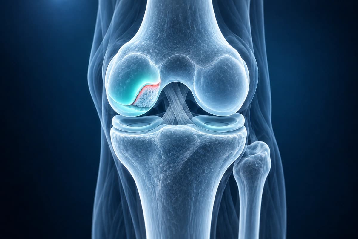
Osteochondritis dissecans (OCD) is a condition where a segment of subchondral bone and its overlying articular cartilage become partially or completely separated from the underlying bone, most commonly affecting the medial femoral condyle. It predominantly occurs in adolescents and young adults, with repetitive microtrauma and vascular insufficiency considered contributing factors.
Common Symptoms
- Activity-related knee pain, often vague and difficult to localize precisely
- Intermittent effusion without a clear traumatic event
- Mechanical symptoms — clicking, catching, or locking — if a fragment has separated
- Tenderness over the affected condyle, classically the medial femoral condyle
- Reduced range of motion and apprehension with condylar palpation during flexion
- Symptoms more common in adolescents and young adults during growth
What We Look For on Imaging
- Well-demarcated subchondral bone fragment on the medial femoral condyle
- High signal interface fluid between the fragment and parent bone on T2 sequences
- Overlying cartilage continuity or disruption assessment
- Cystic changes beneath the fragment indicating instability
- Displaced osteochondral loose bodies within the joint
- Fragment size measurement and percentage of condylar surface involvement
Frequently Asked Questions
How does AI assess OCD lesion stability on MRI?
MRI looks for fluid around or beneath the fragment and cartilage disruption, but stability criteria are less specific in children. AI cannot confirm whether a lesion is stable.
What MRI sequences are most important for osteochondritis dissecans?
Coronal and sagittal T2 fat-saturated sequences are essential for assessing the cartilage surface and the fragment-bone interface.
Can AI predict whether an OCD lesion will heal without surgery?
Healing depends on skeletal maturity, lesion stability, size and symptoms, with follow-up used to assess progress. AI cannot predict whether an individual lesion will heal without surgery.
Sources
- De Smet AA, Fisher DR, Graf BK, Lange RH. Osteochondritis dissecans of the knee: value of MR imaging in determining lesion stability and the presence of articular cartilage defects. AJR Am J Roentgenol. 1990;155(3):549-553.
- Kijowski R, Blankenbaker DG, Shinki K, et al. Juvenile versus adult osteochondritis dissecans of the knee: appropriate MR imaging criteria for instability. Radiology. 2008;248(2):571-578.
- Kessler JI, Nikizad H, Shea KG, et al. The demographics and epidemiology of osteochondritis dissecans of the knee in children and adolescents. Am J Sports Med. 2014;42(2):320-326.
- Cahill BR. Osteochondritis dissecans of the knee: treatment of juvenile and adult forms. J Am Acad Orthop Surg. 1995;3(4):237-247.
Related Articles
Learn when 3T knee MRI can help for cartilage, meniscus, ACL, and subtle bone injuries, and when X-ray, 1.5T MRI, or existing scans are enough.
Learn to understand your knee MRI report, common sequences, and what key findings mean for your diagnosis.
Understand your imaging before your next appointment
MRI, X-ray or CT from a CD, folder or ZIP. A PDF report or photos of the films work too.
Free to open and preview, no account needed. An optional AI report from $10 explains your images in plain language, with questions for your doctor.
Your raw files stay on this device. Only rendered images and minimal context are sent when you request an AI report.
Medical Disclaimer: This page is for informational and educational purposes only. It does not constitute medical advice, diagnosis, or treatment. AI-generated analysis may contain errors. Always consult a qualified healthcare professional for medical decisions. Full Disclaimer