
Meniscus Tear on MRI: Imaging Findings
Meniscal signal grades describe bright signal inside the meniscus on MRI. Grade 1 and grade 2 signal stay inside the meniscus and do not reach its surface; radiologists usually call this intrasubstance degeneration, not a tear. Grade 3 signal reaches the upper or lower surface, and that is the MRI sign of a meniscal tear.
Common Symptoms
- Joint line pain on the inner or outer side of the knee
- Clicking, locking, or catching sensation during movement
- Swelling that develops gradually over 24–48 hours
- Difficulty fully bending or straightening the knee
- Pain when squatting, kneeling, or twisting the leg
- Feeling of the knee giving way under load
What your report says: meniscal signal grade 1, 2 or 3
Report wording you may see
- “Grade 2 intrasubstance signal in the posterior horn of the medial meniscus”
- “Intrasubstance degeneration of the meniscus without a discrete tear”
- “Linear signal contacting the inferior articular surface”
- “Grade 3 signal consistent with a tear”
What the grades do and don't mean
- Grade 1 is a rounded spot of signal and grade 2 a line of signal. In studies comparing MRI with meniscus tissue, both matched degeneration of the tissue rather than a tear.
- Grade 3 signal touches the joint surface of the meniscus. Many radiologists only call a tear when this is seen on at least two images, because a single image is less reliable.
- Meniscal findings become common with age: in a large community study of people aged 50 to 90, most of those with a meniscal tear on MRI had no knee pain in the previous month.
When it tends to matter
Grade 1 or 2 signal on its own is not a tear, so it rarely drives treatment by itself. When grade 3 signal is reported, the tear's pattern, its location and any displaced fragment are the details your doctor weighs together with your symptoms and examination.
Questions for your doctor
- Is the signal grade 2 or grade 3, and on how many images does it reach the surface?
- Which part of the meniscus is affected: the anterior horn, the body or the posterior horn?
- Could this finding explain my symptoms, or is it more likely age-related?
- Would my treatment change if this were called a tear?
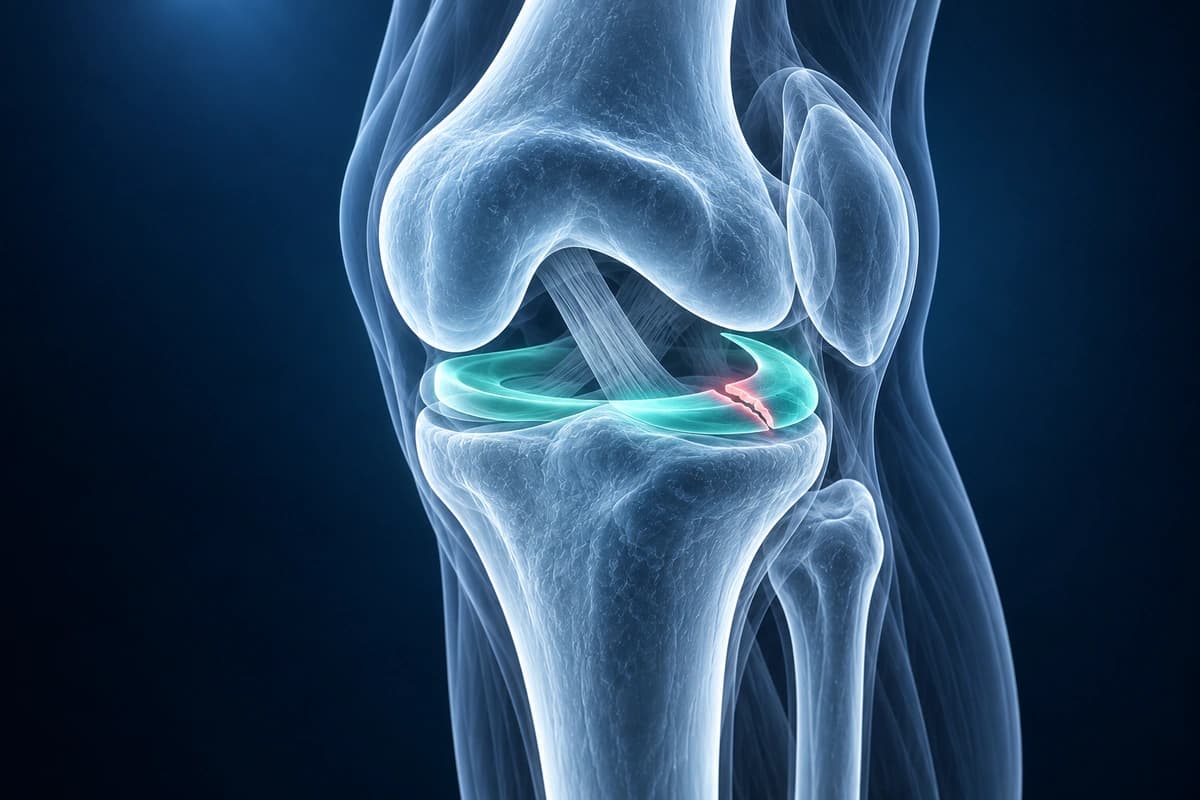
Meniscal tears are among the most common knee injuries, affecting both athletes and older adults. The menisci serve as shock absorbers between the femur and tibia, and tears can cause pain, swelling, and mechanical symptoms.
For how each tear pattern behaves, see meniscus tear types explained; a meniscal tear can also feed fluid into a Baker's cyst behind the knee.
What We Look For on Imaging
- Intrameniscal signal reaching the articular surface on at least two consecutive slices
- Abnormal meniscal morphology (truncation, fragmentation, displaced fragment)
- Bucket-handle tear with displaced meniscal fragment in intercondylar notch
- Radial tear appearing as truncation of meniscal body
- Parameniscal cyst indicating underlying horizontal tear
- Meniscocapsular separation at the periphery
Frequently Asked Questions
Can AI distinguish a radial from a bucket-handle meniscus tear on MRI?
A radial tear crosses meniscal fibers; a bucket-handle tear displaces a longitudinal fragment into the joint, sometimes causing locking. AI cannot reliably classify the tear.
What MRI sequences are most reliable for meniscus evaluation?
Coronal and sagittal proton density fat-saturated (PD-FS) sequences provide the highest contrast for meniscal tissue and intra-substance signal changes.
When does a meniscus tear require surgery?
Surgery is generally indicated for locked knees, bucket-handle tears causing mechanical symptoms, or tears in the avascular zone that will not heal conservatively.
Sources
- Stoller DW, Martin C, Crues JV 3rd, Kaplan L, Mink JH. Meniscal tears: pathologic correlation with MR imaging. Radiology. 1987;163(3):731-735.
- Crues JV 3rd, Mink J, Levy TL, Lotysch M, Stoller DW. Meniscal tears of the knee: accuracy of MR imaging. Radiology. 1987;164(2):445-448.
- De Smet AA, Tuite MJ. Use of the "two-slice-touch" rule for the MRI diagnosis of meniscal tears. AJR Am J Roentgenol. 2006;187(4):911-914.
- Englund M, Guermazi A, Gale D, et al. Incidental meniscal findings on knee MRI in middle-aged and elderly persons. N Engl J Med. 2008;359(11):1108-1115.
- Nguyen JC, De Smet AA, Graf BK, Rosas HG. MR imaging-based diagnosis and classification of meniscal tears. Radiographics. 2014;34(4):981-999.
Related Articles
Learn whether ACL tears can heal on their own, when surgery is needed, and what conservative treatment options exist.
Understand which meniscus tears can heal naturally, the role of blood supply zones, and when surgical repair is recommended.
What ACL reconstruction rehab looks like phase by phase: early goals, key exercises, typical timelines and the tests used before a return to sport.
Understand your imaging before your next appointment
MRI, X-ray or CT from a CD, folder or ZIP. A PDF report or photos of the films work too.
Free to open and preview, no account needed. An optional AI report from $10 explains your images in plain language, with questions for your doctor.
Your raw files stay on this device. Only rendered images and minimal context are sent when you request an AI report.
Medical Disclaimer: This page is for informational and educational purposes only. It does not constitute medical advice, diagnosis, or treatment. AI-generated analysis may contain errors. Always consult a qualified healthcare professional for medical decisions. Full Disclaimer