
ACL Tear Grades Explained: Grade 1, 2, and 3
Understand ACL tear grading from mild sprains to complete ruptures, MRI findings for each grade, and treatment implications.
Key Takeaways
- Grade 1 (sprain): ligament stretched but intact, firm endpoint on Lachman test, 2-4 week recovery
- Grade 2 (partial tear): some fibers disrupted, soft endpoint, treatment is individualized
- Grade 3 (complete tear): all fibers torn, absent endpoint, usually requires surgery in active patients
- MRI is the gold standard for confirming ACL tear grade and identifying associated injuries
- Bone bruising on MRI is present in over 80% of acute complete ACL tears
- Meniscal tears accompany 50-65% of ACL injuries and affect the treatment plan
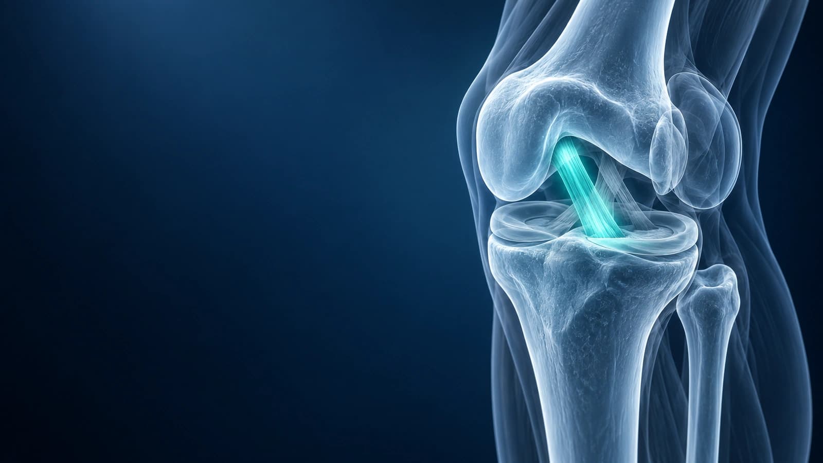
When you receive a diagnosis of an ACL injury, one of the first things your doctor will discuss is the grade of the tear. ACL injuries are classified into three grades based on the severity of ligament damage, and this grading directly influences treatment decisions, rehabilitation protocols, and expected outcomes.
Understanding these grades helps you have more informed discussions with your orthopedic surgeon and know what to expect from your recovery. This guide explains each ACL tear grade in detail, including how they appear on clinical examination and MRI, and what each means for treatment.
For symptoms and treatment options, see the ACL tear overview; if surgery is planned, the ACL recovery timeline explains what each phase involves.
Grade 1 ACL Injury: Ligament Sprain
A Grade 1 ACL injury is a mild sprain where the ligament fibers are stretched but not torn. The ligament remains intact and continuous, and the knee joint retains its structural stability. On clinical examination, the Lachman test and anterior drawer test show a firm endpoint (the knee stops moving at the appropriate point), indicating the ACL is still functional. There may be mild swelling and tenderness, but no sensation of the knee giving way.
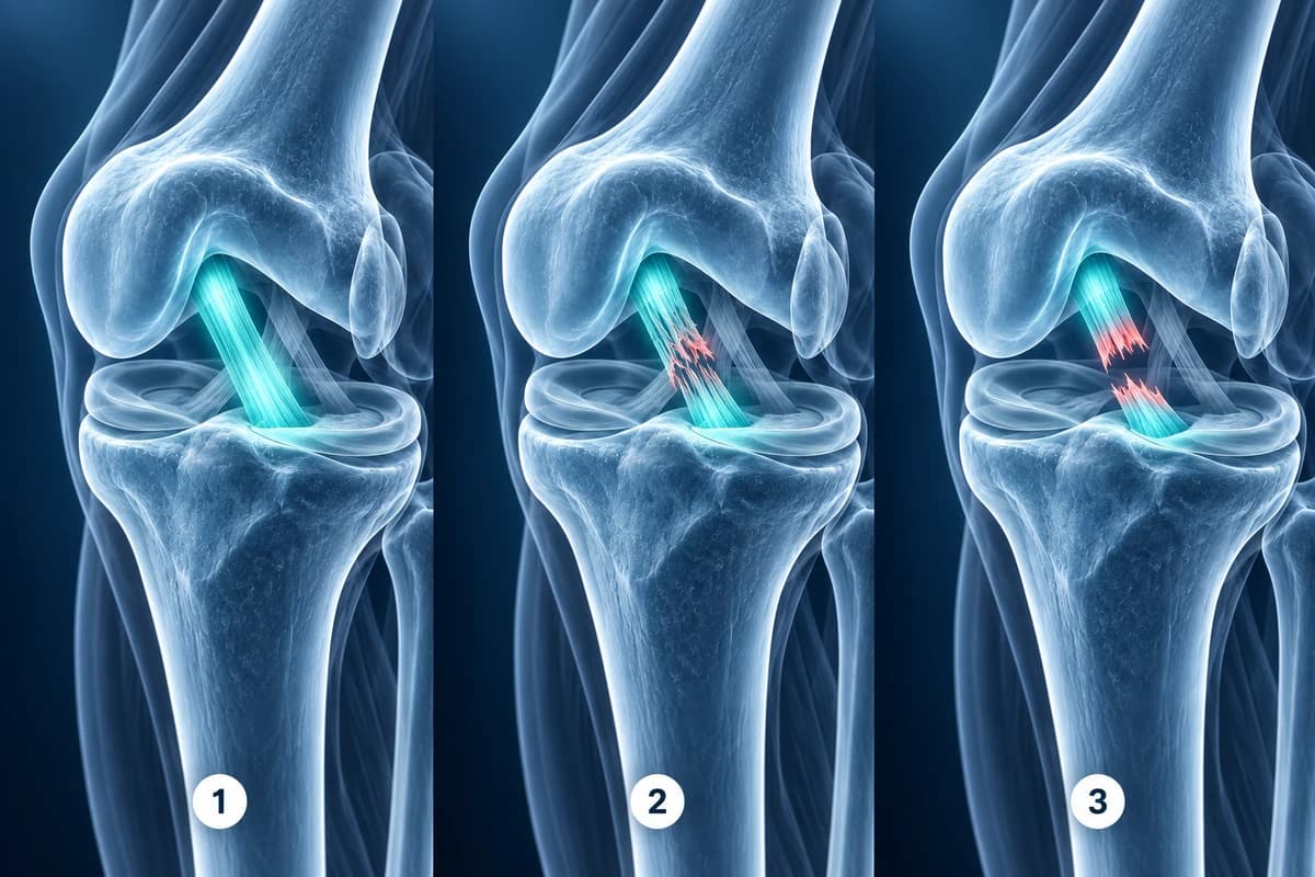
On MRI, a Grade 1 sprain appears as increased signal (brightness) within the ligament on T2-weighted images, indicating edema and microtrauma, but the ligament fibers remain continuous without a visible gap. Treatment is conservative: rest, ice, compression, and elevation followed by physical therapy. Recovery typically takes 2-4 weeks, and most patients return to full activity without restrictions.
Grade 2 ACL Injury: Partial Tear
A Grade 2 ACL injury involves a partial tear of the ligament fibers. Some fibers are disrupted while others remain intact, resulting in partial loss of structural integrity. This is the least common grade — most ACL injuries are either mild sprains or complete tears, with true partial tears accounting for only 10-28% of ACL injuries.
On clinical examination, the Lachman test may show increased laxity compared to the other knee, but with a soft or delayed endpoint. On MRI, a partial ACL tear shows disruption of some ligament fibers with other fibers remaining intact. The overall ligament contour may be irregular or thinned, but a continuous portion can be traced from origin to insertion. Treatment is individualized — many partial tears respond to intensive physical therapy, while patients with persistent instability may benefit from surgical reconstruction or augmentation.
Grade 3 ACL Injury: Complete Tear
A Grade 3 ACL injury is a complete rupture of the ligament. All fibers are torn, and the knee has lost this critical stabilizer. This is the most common ACL injury presentation, often occurring during pivoting, cutting, or landing from a jump, and frequently accompanied by a pop sensation and rapid swelling. On clinical examination, the Lachman test shows significantly increased anterior translation with a soft or absent endpoint.
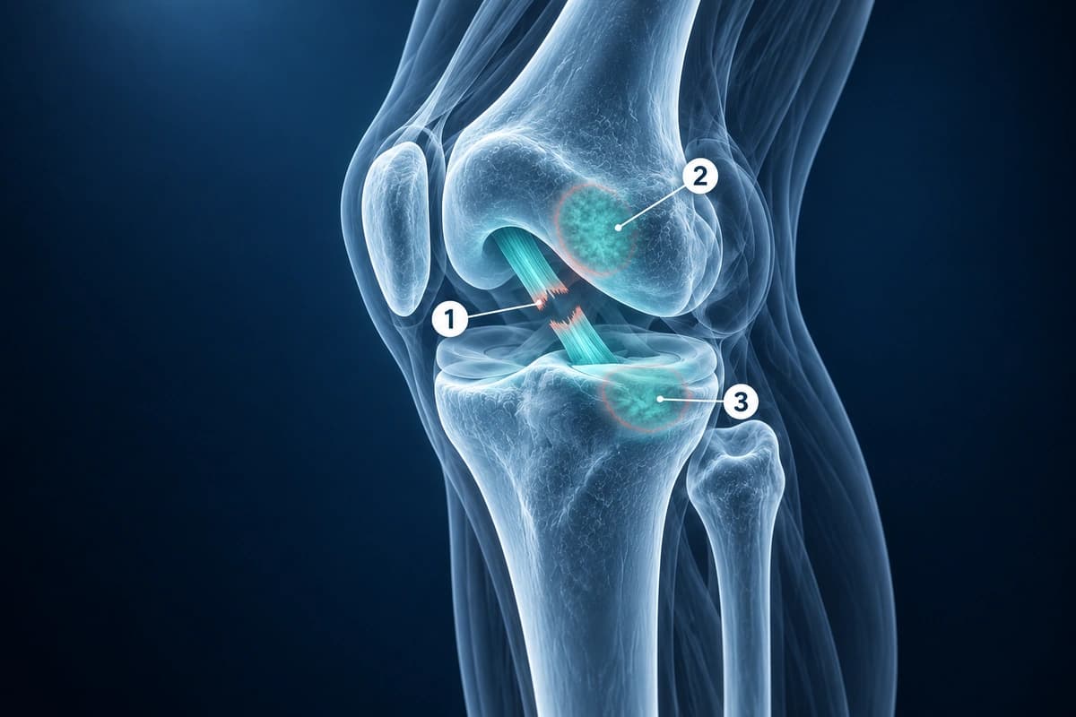
On MRI, a complete ACL tear shows complete discontinuity of the ligament fibers. The ACL may appear as a mass of disorganized, bright signal rather than the normal taut, dark band. Common secondary findings include bone bruising on the lateral femoral condyle and posterolateral tibial plateau (present in over 80% of acute ACL tears), anterior tibial translation, and meniscal tears (present in 50-65% of ACL injuries). For guidance on interpreting these findings, see our article on how to read knee MRI.
Treatment Decisions by Grade
Grade 1 sprains almost always respond to conservative treatment and rarely require surgery. Grade 2 partial tears require individualized assessment — the decision depends on the percentage of fibers torn, the patient's activity level, functional instability symptoms, and the presence of associated meniscal or cartilage injuries. Grade 3 complete tears in young, active patients who participate in pivoting sports are generally recommended for ACL reconstruction.
Older, less active patients or those who do not participate in cutting and pivoting activities may function well without surgical reconstruction (known as copers) with appropriate rehabilitation. For a comprehensive overview of post-surgical rehabilitation, see our article on ACL rehabilitation.
Frequently Asked Questions
Can a partial ACL tear become a complete tear?
Yes. A Grade 2 partial tear can progress to a complete tear if the knee is subjected to further pivoting or twisting forces before the partial tear has healed. Studies suggest that approximately 20-40% of partial ACL tears progress to complete tears, particularly in athletes who return to pivoting sports. This is why activity modification and thorough rehabilitation are essential.
How can my doctor tell the difference between grades on examination?
The Lachman test assesses anterior tibial translation and endpoint quality. Grade 1: firm endpoint. Grade 2: increased translation with soft endpoint. Grade 3: significantly increased translation with no endpoint. MRI provides definitive confirmation.
Do all Grade 3 ACL tears need surgery?
No. While surgery is generally recommended for young, active patients in pivoting sports, some patients with complete ACL tears can function well without reconstruction. These copers typically have strong quadriceps and hamstrings, good proprioception, and modify their activity to avoid high-risk pivoting movements. However, ongoing instability increases the risk of meniscal and cartilage damage over time.
What is the Segond fracture and why is it important?
A Segond fracture is a small avulsion fracture on the lateral tibial plateau, visible on X-ray as a tiny bone fragment pulled away from the tibia. It is considered pathognomonic (uniquely diagnostic) of an ACL tear — when present, it indicates an ACL tear with near 100% certainty. While the fracture itself is small, its identification should prompt MRI to confirm the ACL tear and evaluate for associated injuries.
How accurate is MRI for diagnosing ACL tear grade?
A meta-analysis estimated MRI sensitivity and specificity at 87% and 93% for ACL tears, 89% and 88% for medial meniscal tears, and 78% and 95% for lateral meniscal tears.
Sources
- Noyes FR, Mooar LA, Moorman CT 3rd, McGinniss GH. Partial tears of the anterior cruciate ligament. Progression to complete ligament deficiency. J Bone Joint Surg Br. 1989;71(5):825-833.
- Phelan N, Rowland P, Galvin R, O'Byrne JM. A systematic review and meta-analysis of the diagnostic accuracy of MRI for suspected ACL and meniscal tears of the knee. Knee Surg Sports Traumatol Arthrosc. 2016;24(5):1525-1539.
- Sohn S, AlShammari SM, Hwang BJ, Kim MS. A systematic review of bone bruise patterns following acute anterior cruciate ligament tears: insights into the mechanism of injury. Bioengineering (Basel). 2024;11(4):396.
- Goldman AB, Pavlov H, Rubenstein D. The Segond fracture of the proximal tibia: a small avulsion that reflects major ligamentous damage. AJR Am J Roentgenol. 1988;151(6):1163-1167.
Related Articles
Learn whether ACL tears can heal on their own, when surgery is needed, and what conservative treatment options exist.
Learn to understand your knee MRI report, common sequences, and what key findings mean for your diagnosis.
Related Conditions
Understand your imaging before your next appointment
MRI, X-ray or CT from a CD, folder or ZIP. A PDF report or photos of the films work too.
Free to open and preview, no account needed. An optional AI report from $10 explains your images in plain language, with questions for your doctor.
Your raw files stay on this device. Only rendered images and minimal context are sent when you request an AI report.
Medical Disclaimer: This page is for informational and educational purposes only. It does not constitute medical advice, diagnosis, or treatment. AI-generated analysis may contain errors. Always consult a qualified healthcare professional for medical decisions. Full Disclaimer