
Osteoarthritis on X-ray: Imaging Findings
Knee osteoarthritis (OA) is a degenerative joint disease characterized by progressive cartilage loss, subchondral bone changes, and osteophyte formation. Imaging plays a crucial role in grading severity and monitoring progression. Kellgren-Lawrence grading explained
Common Symptoms
- Gradual onset of deep aching pain worse with activity
- Morning stiffness lasting less than 30 minutes
- Crepitus or grinding sensation during knee movement
- Bony enlargement and loss of full joint range of motion
- Pain that worsens with prolonged standing or stair climbing
- Intermittent joint effusion causing puffiness around the kneecap
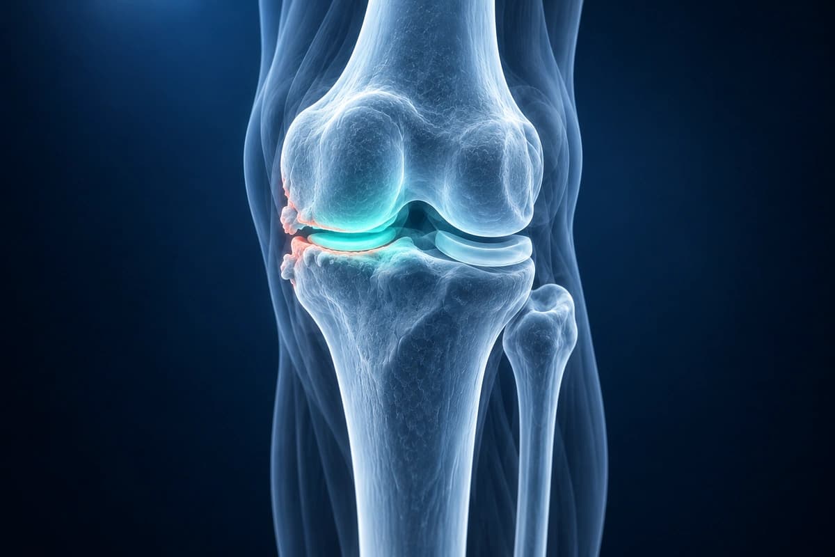
What We Look For on Imaging
- Joint space narrowing on weight-bearing radiographs
- Marginal osteophyte formation at joint margins
- Subchondral sclerosis (increased bone density below cartilage)
- Subchondral cyst formation
- Cartilage thinning or full-thickness loss on MRI
- Bone marrow edema pattern (bone bruise-like changes) on MRI
- Meniscal degeneration and extrusion
Frequently Asked Questions
How is knee osteoarthritis graded on MRI?
Knee osteoarthritis is graded using the Kellgren-Lawrence scale on X-ray or the MOAKS scoring system on MRI. This analysis is for informational purposes only.
Can AI detect early cartilage loss before X-rays become abnormal?
MRI can show cartilage changes before X-rays, but routine osteoarthritis diagnosis usually relies on symptoms and examination. AI cannot confirm early osteoarthritis.
What compartments of the knee does AI evaluate for arthritis?
The knee has medial tibiofemoral, lateral tibiofemoral and patellofemoral compartments; arthritis may affect one or several. AI cannot reliably grade arthritis in these compartments.
Sources
- Hunter DJ, Guermazi A, Lo GH, et al. Evolution of semi-quantitative whole joint assessment of knee OA: MOAKS (MRI Osteoarthritis Knee Score). Osteoarthritis Cartilage. 2011;19(8):990-1002.
- Guermazi A, Niu J, Hayashi D, et al. Prevalence of abnormalities in knees detected by MRI in adults without knee osteoarthritis: population based observational study (Framingham Osteoarthritis Study). BMJ. 2012;345:e5339.
- Katz JN, Arant KR, Loeser RF. Diagnosis and Treatment of Hip and Knee Osteoarthritis: A Review. JAMA. 2021;325(6):568-578.
Related Articles
Understand which meniscus tears can heal naturally, the role of blood supply zones, and when surgical repair is recommended.
Your MRI names a meniscus tear pattern? Learn what horizontal, radial, bucket-handle and complex tears mean, why the zone matters and how each is treated.
Got a Kellgren-Lawrence grade on your knee X-ray report? See what grades 0 to 4 mean, from no arthritis to bone-on-bone, and what each stage implies.
Understand your imaging before your next appointment
MRI, X-ray or CT from a CD, folder or ZIP. A PDF report or photos of the films work too.
Free to open and preview, no account needed. An optional AI report from $10 explains your images in plain language, with questions for your doctor.
Your raw files stay on this device. Only rendered images and minimal context are sent when you request an AI report.
Medical Disclaimer: This page is for informational and educational purposes only. It does not constitute medical advice, diagnosis, or treatment. AI-generated analysis may contain errors. Always consult a qualified healthcare professional for medical decisions. Full Disclaimer