
3D Knee MRI Anatomy Render: How It Works
Learn how compatible knee MRI DICOM slices become a private 3D joint view, what MPR means, and what 3D rendering can and cannot show.

A compatible knee MRI can be rendered as a 3D joint view by stacking DICOM slices into a volume, aligning the image planes, and applying a tissue-sensitive display preset. The 3D view helps orientation and anatomy review, but it does not replace a radiologist's interpretation or clinician review.
The goal is practical: help you understand where structures sit in space before you read the slice-by-slice MRI images and clinician report. It is a viewing aid, not a promise that every ligament, meniscus tear, cartilage defect, or bone bruise will be visible in three dimensions.
What a 3D Knee MRI Render Means
A knee MRI is usually stored as many thin DICOM slices. When the slices belong to a compatible series, the viewer can place them in their original order and treat them as a volume. A 3D render then assigns brightness and transparency to different signal ranges so the joint can be rotated and inspected as a spatial model.
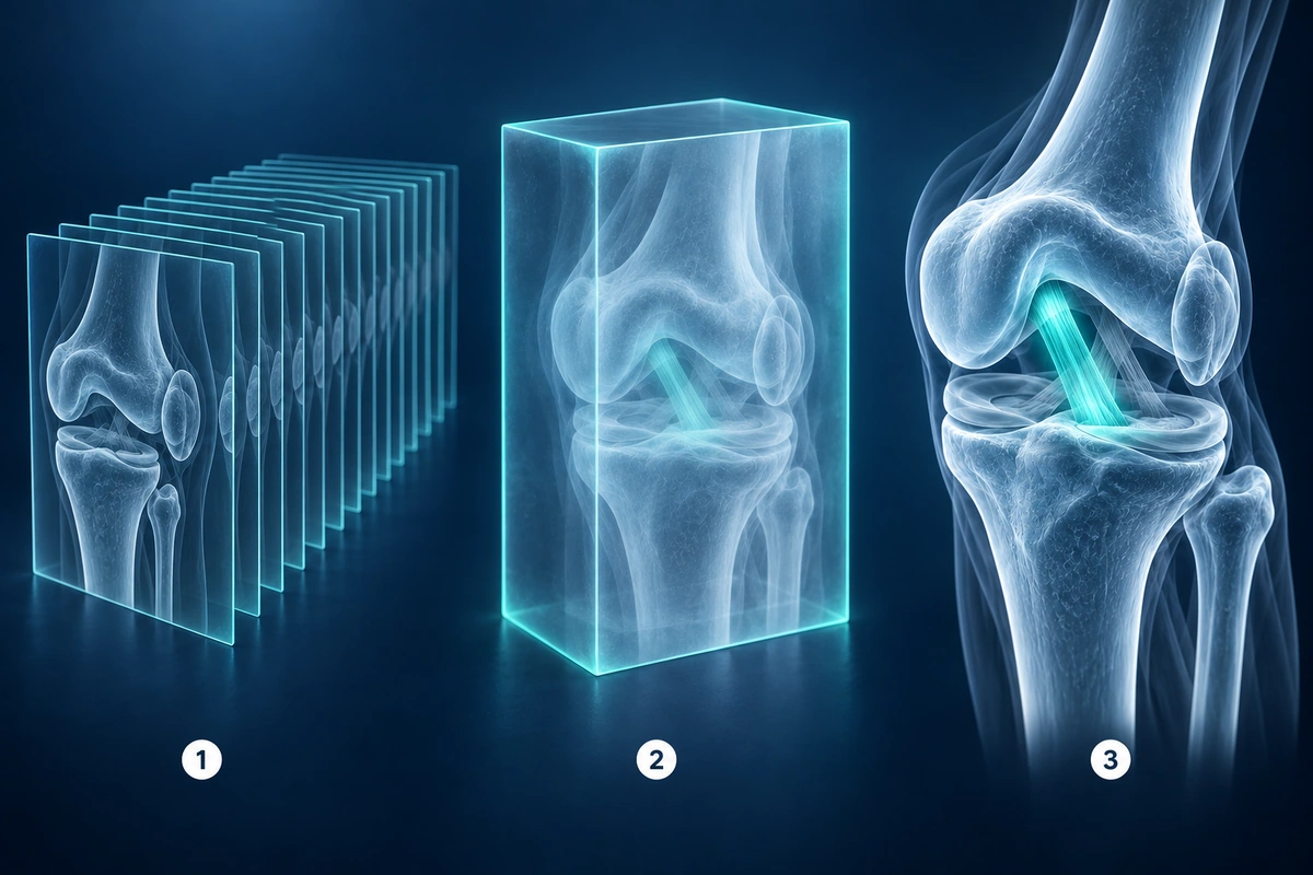
This does not create new medical information. It reorganizes the scan data that is already present. The original 2D images and the radiology report remain the source that clinicians use for interpretation.
MPR vs 3D Volume Rendering
MPR means multi-planar reconstruction. It lets a viewer rebuild the same volume in axial, sagittal, and coronal planes, or sometimes along an oblique angle. MPR is useful when the scan has enough slice coverage and spacing to show anatomy smoothly between the original slices.
3D volume rendering is different. Instead of showing one flat plane at a time, it projects the volume into a rotatable joint view. MPR is usually better for careful slice-by-slice review, while 3D rendering is better for orientation, explaining anatomy, and understanding the relationship between bones and soft tissues.
Viewing Modes Compared
| Mode | Best for | Requires | Limitations |
|---|---|---|---|
| 2D slice view | Careful review of the original MRI sequence | A readable DICOM series or image stack | Orientation can be harder without spatial context |
| MPR | Switching between axial, sagittal, and coronal planes | Volume data with enough slices and consistent spacing | Quality drops when slices are thick, sparse, or misaligned |
| 3D volume render | Orientation, anatomy explanation, and joint overview | Compatible volume data and a useful display preset | Subtle findings may be hidden by the rendering preset |
| AI report explanation | Plain-language support for understanding findings | Rendered images and minimal context selected for analysis | Informational only and not a medical diagnosis |
Which Knee MRI Scans Work Best
3D works best when the study includes a knee MRI series with many closely spaced slices, consistent orientation, and enough signal detail across the whole joint. Dedicated 3D acquisitions and thin-slice sequences usually produce smoother MPR and volume rendering than thick, widely spaced 2D sequences.
Some studies will still load perfectly for regular viewing but will not produce a useful 3D render. A small localizer, a single screenshot, a PDF report, or a sparse series may be viewable as 2D images only. That is a limitation of the source data, not a sign that the scan itself is invalid.
Privacy for the 3D View
For viewing, raw DICOM files are parsed and rendered inside your browser. The 3D knee view is built locally from the compatible image series, so the raw MRI files do not need to be uploaded to a server just to rotate the joint or switch planes.
If you choose AI analysis, the product may send rendered image frames and minimal context needed for explanation, but the raw medical files remain client-side. Avoid sharing identifiable details in free-text notes unless they are necessary for your own clinician discussion.
What the 3D View Cannot Show Reliably
A 3D render can make the knee easier to understand, but it may hide subtle findings. Meniscal signal, partial ligament tears, cartilage fissures, marrow edema, small loose bodies, and postoperative changes often require careful review of the original MRI sequences in multiple planes.

Rendering presets also matter. A preset that makes the bones clear may make soft tissue less visible. A preset that emphasizes fluid may make anatomy look noisy. Treat the 3D view as a companion to the slices, not as the final medical answer.
When to Ask a Clinician
Ask a radiologist, orthopedic specialist, or other qualified clinician to review the MRI if you have pain, swelling, locking, instability, weakness, fever, recent trauma, or symptoms that are getting worse. Imaging needs to be interpreted with your history, physical examination, and treatment goals.
For patient-friendly background on how knee MRI is used and who interprets the exam, RadiologyInfo has a knee MRI guide.
If the 3D render seems to disagree with your report, do not assume one is wrong. The report may describe findings that are not obvious in a surface-style view. Bring your questions to the clinician who can compare the original sequences with your symptoms.
For a broader scan-format view, see the 3D DICOM viewer and MPR guide.
Key Takeaways
- 3D knee MRI rendering is a browser-based viewing aid built from compatible DICOM slices
- MPR shows reconstructed planes, while 3D rendering shows a rotatable volume view
- Thin, closely spaced MRI series usually render better than sparse or thick-slice series
- Raw MRI files stay in the browser for viewing, while optional AI analysis uses rendered images and minimal context
- A 3D view is not a diagnosis and should be reviewed alongside the original images and a clinician's interpretation
Frequently Asked Questions
Can a knee MRI be viewed in 3D?
Yes, some knee MRI studies can be viewed in 3D when they include a compatible DICOM series with enough slices, consistent spacing, and suitable image quality. Other studies may still be useful in the standard 2D slice viewer without producing a good 3D render.
Is 3D MRI rendering the same as a diagnosis?
No. A 3D render is a visualization tool. It can help explain orientation and anatomy, but it does not replace a radiology report, clinician review, physical examination, or medical diagnosis.
Do my MRI files leave my browser for the 3D view?
No. For viewing, the raw DICOM files are parsed and rendered locally in your browser. The 3D view is built from the compatible image series on your device.
Why does 3D work better on some scans than others?
3D rendering depends on the scan protocol. Slice thickness, spacing, field of view, motion, sequence type, and how much of the knee was captured all affect whether the viewer can build a smooth volume.
Related Articles
Understand 3D DICOM viewing, MPR reconstruction, and browser volume rendering for MRI, CT, and CBCT scans, including privacy and scan compatibility limits.
Learn to understand your knee MRI report, common sequences, and what key findings mean for your diagnosis.
Compare browser-based DICOM viewing, cloud upload viewers, and desktop apps so you can open MRI, CT, and X-ray files with the right privacy tradeoffs.
Related Conditions
Ready to analyze your imaging? Upload your MRI or X-ray for AI-powered analysis
Upload your MRI or X-ray DICOM files for private, AI-powered analysis. 4 models analyze independently — raw scan files stay in your browser.
Start AnalysisMedical Disclaimer: This page is for informational and educational purposes only. It does not constitute medical advice, diagnosis, or treatment. AI-generated analysis may contain errors. Always consult a qualified healthcare professional for medical decisions. Full Disclaimer