
ACL Tear on MRI: Imaging Findings
The anterior cruciate ligament (ACL) is one of the most commonly injured knee ligaments, especially in athletes. ACL tears are typically diagnosed through clinical examination and confirmed with MRI imaging. ACL tear grades explained
Common Symptoms
- Sudden pop or giving way during pivoting or landing
- Rapid knee swelling within hours of injury
- Feeling of instability when walking or turning
- Difficulty bearing full weight on the affected leg
- Reduced range of motion, especially full extension
- Pain along the joint line or deep within the knee
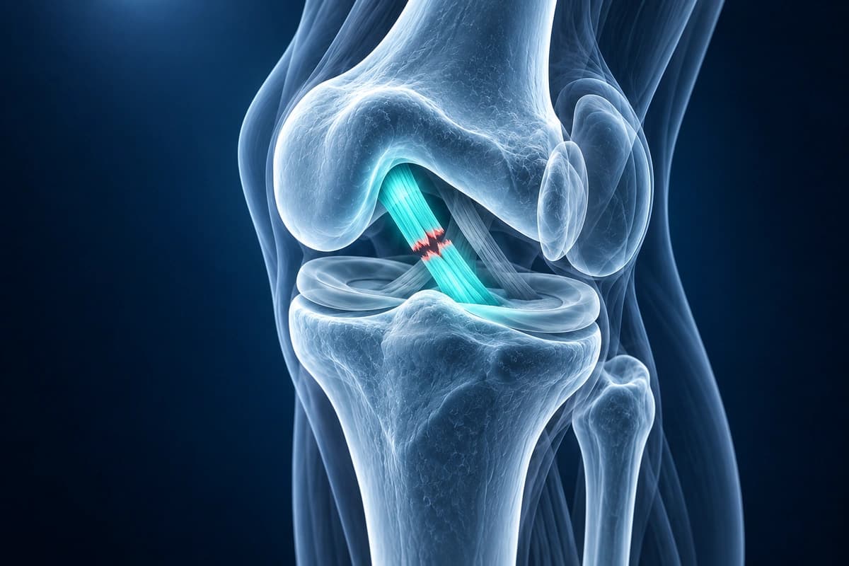
What We Look For on Imaging
- Discontinuity or abnormal signal within ACL fibers on sagittal MRI
- Bone bruise pattern on lateral femoral condyle and posterolateral tibial plateau
- Anterior tibial translation relative to the femur
- Abnormal posterior cruciate ligament (PCL) angle (buckling sign)
- Secondary signs including deep lateral femoral notch sign
- Associated meniscal tears, particularly lateral meniscus posterior horn
Frequently Asked Questions
Can AI detect a partial ACL tear on MRI?
Partial ACL tears can be difficult to distinguish from complete tears on MRI; examination also assesses knee stability. AI cannot confirm whether the remaining fibers function normally.
What MRI sequences are best for ACL evaluation?
Sagittal proton density (PD) and T2-weighted sequences with fat suppression are most useful for ACL evaluation.
How accurate is AI at detecting ACL tears?
Published studies found AI performance comparable to clinicians for ACL tear detection, but prospective validation is still needed. These findings do not establish the accuracy of this service.
Sources
- Tung GA, Davis LM, Wiggins ME, Fadale PD. Tears of the anterior cruciate ligament: primary and secondary signs at MR imaging. Radiology. 1993;188(3):661-667.
- Gentili A, Seeger LL, Yao L, Do HM. Anterior cruciate ligament tear: indirect signs at MR imaging. Radiology. 1994;193(3):835-840.
- Brody JM, Lin HM, Hulstyn MJ, Tung GA. Lateral meniscus root tear and meniscus extrusion with anterior cruciate ligament tear. Radiology. 2006;239(3):805-810.
- Gill SS, Haq T, Zhao Y, et al. AI demonstrates comparable diagnostic performance to radiologists in MRI detection of anterior cruciate ligament tears: a systematic review and meta-analysis. Eur Radiol. 2026;36(4):2500-2517.
Related Articles
Learn whether ACL tears can heal on their own, when surgery is needed, and what conservative treatment options exist.
What ACL reconstruction rehab looks like phase by phase: early goals, key exercises, typical timelines and the tests used before a return to sport.
Understand ACL tear grading from mild sprains to complete ruptures, MRI findings for each grade, and treatment implications.
Understand your imaging before your next appointment
MRI, X-ray or CT from a CD, folder or ZIP. A PDF report or photos of the films work too.
Free to open and preview, no account needed. An optional AI report from $10 explains your images in plain language, with questions for your doctor.
Your raw files stay on this device. Only rendered images and minimal context are sent when you request an AI report.
Medical Disclaimer: This page is for informational and educational purposes only. It does not constitute medical advice, diagnosis, or treatment. AI-generated analysis may contain errors. Always consult a qualified healthcare professional for medical decisions. Full Disclaimer